Hysterosalpingography

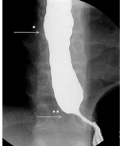
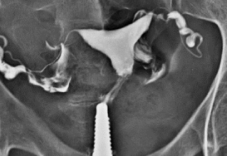
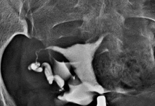
A hysterosalpingography is a type of X-ray that looks at a woman’s uterus (womb) and fallopian tubes (structures that transport eggs from the ovaries to the uterus). This type of X-ray uses a contrast material so that the uterus and fallopian tubes show up clearly on the X-ray images.

The radiologist can watch the dye as it moves through your reproductive system and takes radiographs as the dye opacifies and passes through. The doctor will then be able to see if you have a blockage in your fallopian tubes or other structural abnormalities in your uterus.

Normal
BLOCK –Left side


This test requires that you put on a hospital gown and lie on your back with your knees bent and your feet spread, as you would during a pelvic examination. The radiologist will then insert a speculum into your vagina. This is done so that the cervix, which is located at the back of the vagina, can be seen. You may feel some discomfort.

The radiologist will then clean the cervix .Next, an instrument called a cannula will be inserted into the cervix and the speculum will be removed. The radiologist will insert dye through the cannula, which will flow into your uterus and fallopian tubes.

You’ll then be placed under the X-ray machine, and the radiologist will begin taking X-rays. You may feel some pain and cramping as the dye moves through your fallopian tubes. When the X-rays have been taken, the radiologist will remove the cannula.

You’ll then be prescribed any appropriate medications for pain or infection prevention and you will be sent home after an observation of 30 minutes.


This test is usually ordered  if you’re having trouble getting pregnant or have had pregnancy problems, such as multiple miscarriages.

Hysterosalpingography can help diagnose the cause of infertility.

Infertility may be caused by:

– Structural abnormalities in the uterus, which may be congenital (genetic) or acquired

blockage of the fallopian tubes

-Scar tissue in the uterus

-Uterine fibroids  , tumors or polyps

If you’ve had tubal surgery, a hysterosalpingography  is done to check that this surgery was successful. If you had a tubal ligation (a procedure that closes the fallopian tubes), your doctor may order this test to ensure that your tubes are closed properly. The test can also check that a reversal of a tubal ligation was successful in reopening the fallopian tubes.

FAQ

This test is done ideally in the 1st half of the menstrual cycle  2/3 days after cessation of menses

Ideal time – 7th -10th day

Fasting for 4 hours is preferred.

  • Prior appointment is mandatory
  • An accompanying relative
  • Doctors referral paper if available ( Not mandatory )
  • Exact date of last menses
  • â–ŞIn case of missed period a recent Negative UPT report is mandatory       

Reports are issued the same day / next day

Mailing / Whats ap on request

After the test, you may continue to have cramps similar to those experienced during a menstrual cycle. You may also experience vaginal discharge or slight vaginal bleeding. You should use a pad instead of a tampon to avoid infection during this time.

Some women also experience dizziness and nausea rarely vomiting during or after the test. These side effects are normal and will eventually go away.

However, let your doctor know if you experience symptoms of infection, including:

  • Fever  
  • Severe pain and cramping       
  • Foul-smelling vaginal discharge  
  • Heavy vaginal bleeding  

Complications from a hysterosalpingography are rare. Possible risks include :

  • Allergic reaction to contrast dye
  • endometrial (uterine lining) or fallopian tube infection
  • injury to the uterus, such as perforation
  • Pregnancy
  • Pelvic inflammatory disease
  • Unexplained vaginal bleeding

Schedule Your Appointment

Test timingPatient PreparationPre-procedure MedicationTest durationCommon IndicationsRisks and complications
HSG (Hysterosalpingography)Anytime during the day, 7th to 10th day of menstrual cycle, Prior appointment requiredFasting for 2-3 hrs, Inform the doctor about allergies to any medicines, latex, or anesthetics and about existing medical conditions. Remove all jewelry and aids or devices such as dentures, hair clips etc; and wear a hospital gown. Let your doctor know about all the medicines you are currently taking, Carry all your relevant old reports and show it to the doctor before the testProphylactic antibiotics before or after the test to prevent infection; SOS Atropine inj IV /IM and a Tetanus toxoid injection30 minsInfertility, Diagnose fallopian tube blockage, Adhesions in uterus, After tubal ligation / tubal ligation reversalRare. Possible risks include- Allergic reaction to contrast dye, Endometrial (uterine lining) or fallopian tube infection, Injury to the uterus, such as perforation

BARIUM STUDIES

A barium X-ray is a radiographic (X-ray) examination of the gastrointestinal (GI) tract. Barium X-rays (also called upper and lower GI series) are used to diagnose abnormalities of the GI tract, such as tumors, ulcers and other inflammatory conditions, polyps, hernias, and strictures.

Barium is a dry, white, chalky powder that is mixed with water to make barium liquid. Barium is an X-ray absorber and appears white on X-ray film. When instilled into the GI tract, barium coats the inside wall of the esophagus, stomach, large intestine, and/or small intestine so that the inside wall lining, size, shape, contour, and patency (openness) are visible on X-ray. This process shows differences that might not be seen on standard X-rays.

Barium is used only for diagnostic studies of the GI tract.

BARIUM SWALLOW
Barium FOLLOW THROUGH

FAQ

This test is done any day of the month.

  • Fasting for 4 hours is needed for a barium swallow.
  • Overnight fasting and good bowel clearance needed for a Follow-through
  • We prescribe laxatives when required at the time of appointment

Test timingPatient PreparationTest durationCommon IndicationsRisks and complications
Barium SwallowAnytime during the day, Prior appointment requiredFasting for 2 hrs, Inform the doctor about allergies to any medicines, latex, or anesthetics and about existing medical conditions. Remove all jewelry and aids or devices such as dentures, hair clips etc; and wear a hospital gown. Let your doctor know about all the medicines you are currently taking, Carry all your relevant old reports and show it to the doctor before the test30 minsDifficulty in swallowing, Gastro-oesophageal reflux, Hiatus hernia, Oesophageal spasmConstipation or impacted stool, Trouble with bowel movements, Inability to have a bowel movement or pass gas, Pain or swelling of the abdomen, Allergic reaction or anaphylaxis to the barium drink, Barium aspiration in the windpipe
Barium Follow-throughEarly morning, Prior appointment requiredOvernight fasting, Laxatives ( 2 tabs Dulcoflex)previous night for bowel clearance, Inform the doctor about allergies to any medicines, latex, or anesthetics and about existing medical conditions., Remove all jewelry and aids or devices such as dentures, hair clips etc; and wear a hospital gown. Let your doctor know about all the medicines you are currently taking, Carry all your relevant old reports and show it to the doctor before the test2-4 hrsConstipation or impacted stool, Trouble with bowel movements, Inability to have a bowel movement or pass gas, Pain or swelling of the abdomen, Allergic reaction or anaphylaxis to the barium drink

  • Prior appointment is mandatory
  • An accompanying relative
  • Doctors referral paper if available (Not mandatory)

Reports are issued the same day / next day

Mailing / WhatsApp on request

IVP - Intravenous Urography

An intravenous pyelogram, also called an excretory urogram, is an X-ray exam of your urinary tract.

An intravenous pyelogram lets your doctor view your kidneys, bladder and the tubes that carry urine from your kidneys to your bladder (ureters).

An intravenous pyelogram may be used to diagnose disorders that affect the urinary tract, such as kidney or ureteric stones, bladder stones, enlarged prostate, kidney cysts or urinary tract tumors.

During an intravenous pyelogram, an X-ray dye (iodine contrast solution) is injected into a vein in the arm. The dye flows into your kidneys, ureters and bladder, outlining each of these structures. X-ray pictures are taken at specific times during the exam, so the doctor can clearly see your urinary tract.

FAQ

This test is done any day of the month.

  • Overnight fasting and good bowel clearance needed
  • We prescribe laxatives when required at the time of appointment
  • Prior appointment is mandatory
  • An accompanying relative
  • Normal Blood Creatinine report done within last 2 days
  • Referral  paper of doctor
  • Previous USG / X Ray / CT Scan reports

Reports are issued the same day / next day

Mailing / WhatsApp on request

Schedule Your Appointment

Request An Appointment

To schedule an appointment please contact our office by phone or by filling in the form below and we will get back to you